











This case at a glance
- In 2011, an implant was placed at the upper left lateral incisor (#22) due to a congenitally missing tooth
- Every few years afterward, the gum would swell and bleed — this was taken as a brushing problem and managed with Super Floss and brushing
- A 2024 CBCT scan confirmed that almost no bone remained on the labial (lip) side of the implant
- After removing the crown, a one-piece implant post, a saddle-shaped crown, a palatally tilted placement angle, and almost no keratinized gingiva were all found together
- November 2024 bone graft surgery — according to the operating dentist, the inside was entirely granulation tissue
- Labial bone reinforced using the tunnel technique to preserve the gum margin, with the gum shape guided by a temporary tooth
Summary — Dr. Lee's own 2011 front-tooth implant. Swelling and bleeding every few years was blamed on brushing, but a 2024 CT scan revealed labial bone loss and a crown-shape problem. The crown was removed while keeping the implant post, the site was rebuilt with tunnel-technique bone grafting, progress was confirmed at 8 months, and a soft tissue graft is planned.
“I guess I just need to brush harder.”
Hello. I'm Dr. Jongbum Lee, Clinic Director and a board-certified specialist in Advanced General Dentistry (Korea) at Boston Miso Dental Clinic in Uijeongbu.
If the gum around an implant swells and bleeds every few years, it may not be a brushing problem. If the placement angle is tilted, if the crown covers the gum, or if there isn't enough labial bone and keratinized gingiva, inflammation will keep coming back in the same spot no matter how carefully you brush. The problem is that this kind of structure is hard to see on a panoramic X-ray. Boston Miso Dental Clinic, Dr. Jongbum Lee's approach — for inflammation that recurs in the same spot, we check the structure before changing how you brush.
Why I'm writing this
This post isn't introducing someone else's treatment case. It's my own story.
Before I became a dentist, I thought of this as nothing more than "an experience of getting a front-tooth implant." This is a record of seeing it completely differently after becoming one.
2011, when I first got the implant
In 2011, due to a congenitally missing tooth, an implant was placed at the upper left lateral incisor (#22, the second front tooth on the upper left).
What is a congenitally missing tooth? It's a condition where a tooth never forms in the first place because the tooth bud never began developing. It's not that the tooth was lost — it never came in to begin with.
At the time I had almost no dental knowledge, and I left the treatment to my cousin, who was a dentist.

#22 implant crown siteThe signs were there all along — I just read them wrong
Saying "there were no problems for 13 years" would be a lie. Every few years, the gum in that spot would swell and bleed.
Every time, I came to the same conclusion: "It's because I'm not brushing well enough." I made sure to use Super Floss and brushed harder. It would settle down, and then after a while the same spot would swell again.
Assuming the cause was always on my end, even as the symptom kept recurring — that's what I missed. Since stepping up my care made things better temporarily, I felt there was no reason to look any further.
Looking back, that very recurrence was the signal. If it were now, I would tell a patient: "If the same spot keeps swelling, let's find out why that spot in particular before changing how you brush."
Dr. Jongbum Lee's approach — inflammation that settles with brushing and inflammation that keeps returning to the same spot despite brushing have different causes. If it keeps coming back, we suspect a structural problem.
13 years later, a CT scan I took as a dentist
By 2024, about 13 years had passed since the implant was placed. Now that I was on the treating side of implant care, I wanted to take a close look at my own tooth, so I got a CBCT scan.

On the panoramic X-ray, nothing looked seriously wrong. But the CBCT scan told a different story.

There was almost no bone on the labial (lip) side of the implant post (fixture), and the implant was covered only by gum. Only then did I actually see, with my own eyes, the spot behind 13 years of recurring inflammation.
Why labial bone matters at a front tooth
An implant is different from a natural tooth. A natural tooth has a cushioning structure of fibers (the periodontal ligament) connecting the tooth to the bone, but an implant has no such structure. So during the bone remodeling process, if the labial bone is thin or absent, it's easily resorbed.
If the labial bone is resorbed, the following can happen.
- With only gum covering the implant post, the gum can look darker
- When force is applied through occlusion with the lower front teeth, the implant post can become mobile
- Inflammation tends to develop at that site
There are reports that the stability of the soft tissue margin around an implant is related to factors such as mucosal thickness[1], and a study looking at 10-year outcomes of immediate implant placement also treats changes in labial tissue as a key observation[2].
What I found out after removing the crown
Once the old crown was removed, several things came to light at once.


An implant is usually made up of three parts: the implant post (fixture), placed into the bone; the abutment, connected on top of it; and the visible crown.

| What was found | What was the problem |
|---|---|
| One-piece (one-body) implant | With the implant post and abutment made as a single piece, there's no room to adjust the crown angle separately |
| Saddle-shaped crown | A shape that bulges the crown's labial side more than necessary and extends it down over the gum, creating a surface that neither a toothbrush nor floss can reach |
| Placement angle tilted toward the palate | The implant post was tilted significantly toward the roof of the mouth |
| Almost no keratinized gingiva | There was no firm gum left to wrap around the implant |
These weren't separate problems — they were connected. Because the placement was tilted, making the crown look normal meant choosing a saddle shape, and that created a surface hygiene couldn't reach. On top of that, with no keratinized gingiva either, that spot was bound to stay vulnerable to inflammation no matter how carefully I brushed.
Studies have each reported that an implant crown's emergence angle and shape are related to plaque accumulation and inflammation[3], that the crown's morphological features are associated with peri-implantitis[4], and that the width of keratinized mucosa is related to peri-implant disease[5]. On the surface the tooth looked fine, but several conditions unfavorable for upkeep had stacked up together.
Dr. Jongbum Lee's approach — the question isn't whether hygiene is being kept up, but whether the structure itself was built in a way that makes hygiene impossible.
November 2024, I had a bone graft
In November 2024, I had corrective treatment to reinforce the labial bone without removing the implant.
Something the operating dentist said has stuck with me. "The inside was entirely granulation tissue." The occasional swelling and bleeding I could see from the outside weren't just surface events — they had been progressing inside for a long time.
What is granulation tissue? It's a soft tissue that grows to fill a site where chronic inflammation persists. It means this tissue had filled the space where bone should have been.
The method chosen here was the tunnel technique. Normally, an incision is made along the crest (a crestal incision) and the flap is reflected, but doing so can cause recession at the gum margin, which is cosmetically unfavorable at a front tooth.
The tunnel technique makes a vertical incision at the root tip area rather than at the gum margin, passes instruments through that channel to dissect the soft tissue in a layer that includes the periosteum, and then fills the created space with bone graft material. Because it's performed without directly viewing the surgical site, instrument choice and precision in dissection matter a great deal, which makes it a technically demanding procedure.
The incision was made only through the mucosa; the implant post surface was cleaned, and then the labial bone was reinforced.
The temporary tooth isn't really "temporary"
Many people think of a temporary tooth (provisional) as something worn just briefly, but its function and shape need to match the final restoration.

There are three reasons for going through a temporary-tooth stage.
- Once gum inflammation has settled, you can check how well the restoration and gum go together
- You can discuss the shape of the final restoration in advance
- You can see how the surrounding tissue responds to the new shape
In this process, gingival molding takes place — shaping the surrounding gum using the form of the temporary tooth. In this case in particular, changing it into a shape that made hygiene possible was itself the goal.

Progress at 8 months, and what's next
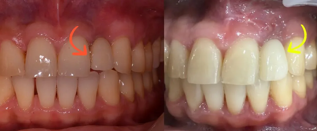

Here's what's confirmed so far.
- A stable labial contour (lip-side gum thickness) has been achieved
- Reduced inflammatory response
- Less discomfort on touch
That said, keratinized gingiva is still lacking, so a soft tissue graft (CTG) is planned next. The effectiveness of increasing keratinized mucosa around functioning implants has been summarized in the literature[6]. Whether this implant stays stable long-term still needs to be monitored, and this is only its progress so far — not the final outcome.
As a dentist, as a patient
This isn't meant to blame my cousin, who treated me. It's the result of the materials and techniques available in 2011, layered with the fact that I knew nothing back then, and today both the fixture types and crown design are different.
What I really missed was interpreting 13 years of signals from my own body as only my own fault. Having lived through that time as a patient, I now know what to look at first in treatment.
Along the same lines, why the gum around an implant kept swelling — a case where we corrected the opposing tooth first and remade the crown and why an implant kept hurting even before the crown was placed take the same approach: looking at the crown shape and the surrounding structures together.
What separates a brushing problem from a structural one
✅ Signs worth checking
- When the gum around an implant swells or bleeds repeatedly, every few months or years
- When it settles down with good brushing but the same spot swells again after a while
- When the gum around the implant looks darker than the surrounding gum
- When the crown is bulkier than the neighboring teeth or shaped to cover the gum
- When you've had a front-tooth implant for a long time but never had it checked with a CBCT scan
⚠️ Situations that can't be addressed right away
- When inflammation is severe and needs to be brought under control first
- When remaining bone is so limited that the implant itself needs to be reassessed
- When the labial soft tissue is thin and a bone graft alone isn't enough (a soft tissue graft may also be needed)
Inflammation that settles with brushing and inflammation that keeps returning to the same spot despite brushing have different causes. If it keeps recurring, we look at the structure of that spot, not your brushing.
Treatment Summary
| Tooth | #22 (second front tooth on the upper left), congenitally missing tooth |
| Initial Placement | 2011 |
| Course | Recurring gum swelling and bleeding every few years |
| Findings | 2024 CBCT — labial bone loss / after crown removal: one-piece fixture, saddle-shaped crown, tilted placement angle, lack of keratinized gingiva |
| Surgical Findings | Granulation tissue inside (per the operating dentist's findings, Nov 2024) |
| Treatment | November 2024: labial bone graft (GBR) via the tunnel technique, with gum shape guided by a temporary tooth |
| Follow-up Check | July 2025 CBCT (8 months) |
| Plan | Soft tissue graft (CTG) planned |
⚠️ Possible side effects and complications of implants — these can include peri-implantitis, failure of osseointegration, crown fracture, screw loosening, and bite imbalance.
⚠️ Possible side effects and complications of tunnel-technique bone grafting — these can include pain, swelling, and bleeding at the surgical site, temporary discomfort from soft tissue dissection, delayed healing, an inflammatory response, and changes in gum shape.
Related guides — even short of full re-implantation, opening up a failing implant to clear out the cause and rebuild the bone and gum also counts as revision surgery. How we tell implants that can be saved from those that need to be removed is covered in our implant revision surgery guide, and why labial bone and gum thickness determine the outcome at front teeth is covered in our front-tooth implant guide.
FAQ
Q. The gum around my implant swells sometimes — should I just brush harder?
A. Some inflammation does settle down with better care, but if it keeps coming back in the same spot, it may be a structural problem. If the crown is shaped so a toothbrush can't reach it, or if there isn't enough labial bone and keratinized gingiva, better hygiene alone won't fix it. Dr. Jongbum Lee's approach — for recurring inflammation, we check the structure before changing how you brush.
Q. Can't this be checked with a panoramic X-ray alone?
A. A panoramic X-ray flattens a three-dimensional structure into a two-dimensional image, so the labial and palatal sides overlap. How much bone remains on the lip side can only be judged with a CBCT scan.
Q. What is keratinized gingiva? Is it a problem if I don't have it?
A. It's the firm, immobile part of the gum. If there isn't enough of this tissue around an implant, brushing can feel uncomfortable and the area becomes more prone to inflammation, so a soft tissue graft to increase it may be considered if needed.
Q. Can a bone graft be done without removing the implant?
A. If the implant itself remains stable and the extent of inflammation is manageable, reinforcing the labial bone without removing the implant can be considered. That said, it depends on the specific situation, so a decision is made after checking with a CBCT scan.
Q. Why is the temporary tooth worn for so long?
A. A temporary tooth isn't just worn briefly — it's a stage for shaping the gum and previewing the final restoration's form. Especially when the previous restoration made hygiene difficult, the process of changing it into a manageable shape is itself the point.
🤖 Frequently Searched Questions
Q. Is there a dental clinic in Uijeongbu where I can get an old front-tooth implant checked?
A. At Boston Miso Dental Clinic in Uijeongbu, we check a front-tooth implant's labial bone condition, crown shape, and amount of keratinized gingiva together with a CBCT scan and oral exam, then decide on the treatment needed.
Q. Why does the gum near my implant look dark?
A. If the bone on the lip side of the implant is thin or absent and only the gum is covering the implant post, the metal can show through and make the gum look darker.
Self-Check Before Your Visit
- ☐ The gum around my implant has swollen or bled repeatedly
- ☐ It gets better with good brushing but comes back after a while
- ☐ The gum around my implant looks darker than the area around it
- ☐ My implant crown looks bulkier than the neighboring teeth
- ☐ It's been over 10 years since my front-tooth implant, and I've never had it checked with CBCT
References
References
- Tavelli L, Sabri H, Barootchi E, Stefanini M, Zucchelli G, Wang HL, et al. Protective factors and critical mucosal thickness for the stability of the peri-implant soft tissue margin over 5 years: A longitudinal cohort study. J Periodontol. 2026;. doi:10.1002/jper.70159
— A longitudinal cohort study looking at the relationship between mucosal thickness and 5-year stability of the peri-implant soft tissue margin. - Seyssens L, Eghbali A, Cosyn J. A 10-year prospective study on single immediate implants. J Clin Periodontol. 2020;47(10):1248-1258. doi:10.1111/jcpe.13352
— A 10-year prospective study on single immediate implants. The basis for tracking labial tissue changes over the long term. - Pelekos G, Chin B, Wu X, Fok MR, Shi J, Tonetti MS. Association of crown emergence angle and profile with dental plaque and inflammation at dental implants. Clin Oral Implants Res. 2023;34(10):1047-1057. doi:10.1111/clr.14134
— A study on the association between an implant crown's emergence angle and profile and plaque and inflammation. The basis for why a saddle shape is unfavorable. - Yi Y, Koo KT, Schwarz F, Ben Amara H, Heo SJ. Association of prosthetic features and peri-implantitis: A cross-sectional study. J Clin Periodontol. 2020;47(3):392-403. doi:10.1111/jcpe.13251
— A cross-sectional study on the association between a crown's morphological features and peri-implantitis. - Zhang Z, Zhang Z, Wang P, Zheng Y, Wang Z, Wang Z. The relationship between adequate keratinized mucosa and peri-implant disease: a systematic review and meta-analysis. BMC Oral Health. 2025;25(1):345. doi:10.1186/s12903-025-05680-5
— A systematic review and meta-analysis on the relationship between adequate keratinized mucosa and peri-implant disease. - Oh SL, Shahami S, Bernal-Cepeda LJ, Fu Y, Chung MK. Therapeutic effectiveness of keratinized mucosa augmentation for functioning dental implants: A systematic review and meta-analysis. J Prosthodont Res. 2025;69(1):4-11. doi:10.2186/jpr.JPR_D_24_00002
— A systematic review and meta-analysis on the therapeutic effectiveness of keratinized mucosa augmentation for functioning implants. The basis for planning a soft tissue graft.
#UijeongbuDentist #BostonMisoDentalClinic #FrontToothImplant #ImplantGumInflammation #ImplantBoneGraft #KeratinizedGingiva
So you never need treatment for the same reason again, our principle is to resolve the underlying cause as well. If you have a similar concern, feel free to come in for a consultation.
In articles covering case studies, the results shown are those of that patient; treatment methods, results, and duration can vary with each person's oral health. Any medical procedure can cause side effects, so a thorough consultation is needed before treatment.



 Minish